

Este kit está diseñado utilizando la tecnología de integración del método de Golgi-Cox1,3 de Ramón- Moliner4, Glaser and Van der Loos2. En el kit, la tecnología se simplifica y se cambió, sino que también mejoró de una manera que los mismos resultados se obtienen con el protocolo simple.
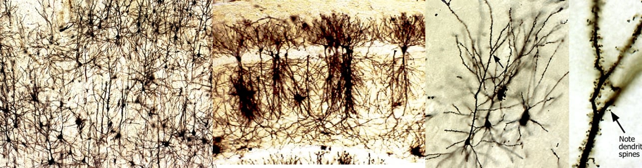
El método de Golgi-Cox1,3 es la técnica más eficaz para examinar la morfología normal y anormal de las neuronas, y la glía.
Cada kit contiene:
Solution A 250 ml
Solution B 250 ml
Solution C 250 ml x 2
Solution D 250 ml
Solution E 250 ml
Glass Specimen Retriever 2
Natural hair paintbrush 2
Dropping bottle 1
User Manual 1
Podría ver los productos que ofrecemos:
Referencia |
Producto |
Cantidad |
Precio |
| PK401 | FD Rapid GolgiStain Kit (large) | kit para 50 cerebros del ratón | 733 € |
| PK401A | FD Rapid GolgiStain Kit (small) | kit para 25 cerebros del ratón | 575 € |
| PK401-C | FD Rapid GolgiStain kit – Solution C | 250 ml | 189 € |
| SS201 | Tissue preparation & Golgi-Cox Staining with FD Rapid GolgiStain™ Kit | un kit | Pregúntanos |
Guarda todos los kits abajo al temperatura ambiental.
")
|
|
||||||
|
|
|
||||||
|
|
|
||||||
En stock: Sí;
Podría descargar el manual para que viera todo el procedimiento de tinción del Golgi Stain:
- Corsi P. (1987) Camillo Golgi’s morphological approach to neuroanatomy. In Masland RL, Portera-Sanchez A and Toffano G (eds.), Neuroplasticity: a new therapeutic tool in the CNS pathology, pp 1-7. Berlin: Springer.
- Glaser ME, and Van der Loos H. (1981) Analysis of thick brain sections by obverse-reverse computer microscopy: application of a new, high clarity Golgi-Nissl stain. J. Neurosci. Meth. 4:117-25.
- Ramón-Moliner E. (1970) The Golgi-Cox technique. In Nauta WJH and Ebbesson SOE (eds.), Contemporary Methods in Neuroanatomy. pp 32-55, New York: Springer.